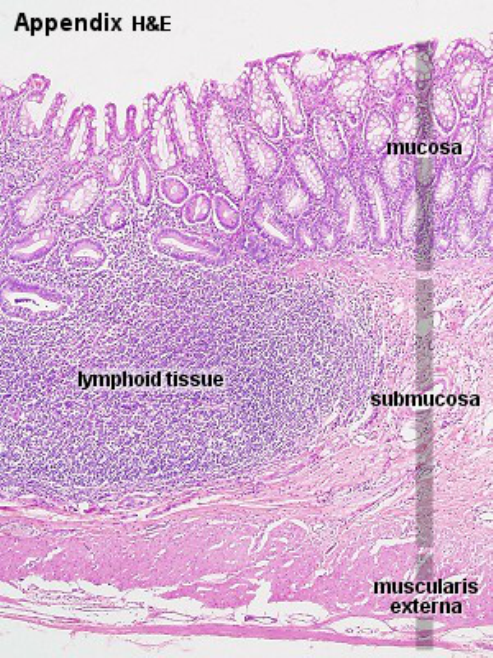
GUT-Associated Lymphoid Tissue
The GI tract is the largest immune organ in the body, containing 65% of immune tissue overall and up to 80% of the immunoglobulin-producing tissues of the body.
Gut-associated lymphoid tissueis characterized by
|
These two functional hallmarks of the GALT are representative of the fact that the intestine is a very large immunologic organ in addition to its obvious other functions associated with digestion and endocrine secretion.
Its functions are organized anatomically and include two components. The first component is the so-called organized GALT, which includes the Peyer patches, isolated lymphoid follicles, and lymphocyte-filled villi. The second component, the so-called diffuse GALT, comprises anatomic structures that are diffusely contained within the lamina propria.
The mucosal-associated lymphoid tissues is regulated state of controlled (or physiologic) inflammation.
Thus, the gut is poised for, but actively restrained from, full action and notable for a tendency to suppress responses, a characteristic referred to as oral tolerance.
Oral tolerance and controlled inflammation are unique hallmarks of the GALT.
Both manifestations of the tendency of mucosal-associated lymphoid tissues to tightly regulate immune responses.
The normal motility, villous microanatomy, rich blood supply, and epithelial intercellular tight junctions contribute to the overall integrity and barrier function of the GI tract.
In response to luminal nutrients, propulsive contractions assist in controlling the concentration of luminal bacteria, and the secretion of bile salts, mucus glycoproteins, and secretory IgA retard bacterial adhesion to gut epithelial cells and subsequent translocation.3,4
The healthy gut acts as an important antigen-sensing organ, in which bacterial antigen is sampled and processed by the M cells, ultimately stimulating the release and maturation of a population of pluripotential stem cells or naïve CD4 helper T lymphocytes.5,6
+++
Organized GALT
The organized GALT structures are often characterized by specialized types of epithelium such as the so-called microfold cell (M cell). M cells are unique epithelial cells that overly the Peyer patches, with its rich content of associated lymphocytes and dendritic cells that allow for selective uptake of and response to distinct types of antigens. The Peyer patches and other organized lymphoid structures are distributed throughout the gastrointestinal tract but are especially congregated in the distal ileum. They are mainly inductive sites where antigens, including bacterial antigens, are taken up, processed, and presented by dendritic cells for the education of the GALT-associated lymphocytes.
The diffuse GALT constitutes the majority of lymphoid tissue within the small and large intestine and is characterized by a single layer of simple, columnar epithelial cells that separates the lumen of the intestines from the lamina propria. This epithelium and its associated intraepithelial lymphocytes, and underlying scattered lymphoid and dendritic cells are a major effector site for protecting the large surface of intestines that must be defended from epithelial exposures to pathogens. In addition, this epithelial surface is highly responsible for participating in the maintenance of a regulated immune response to the wide variety of microbes associated with the normal commensal microbiota.
Both the epithelial compartment above the epithelial basement membrane and the subepithelial compartment below the epithelial basement membrane that is contained within the lamina propria participate in the two major arms of the immune system. These are the innate and adaptive (or specific) immune systems (Table 2–2).
Table 2–2. Innate and adaptive immunity.
| Type of Immunity | Receptor | Ligand | Cell Type |
|---|---|---|---|
| Innate immunity (rapid, hard wired) | TLR2 | Peptidoglycan | Macrophages |
| TLR4 | LPS | Macrophages/IEC | |
| TLR5 | Flagellin | Macrophages/IEC | |
| TLR9 | Bacterial DNA | Dendritic cells | |
| CRP | Bacterial carbohydrate | Serum | |
| NOD2 | Muramyl dipeptide | Dendritic cells/IEC | |
| Adaptive or specific immunity (delayed with memory) | TCR | HLA plus peptide | T cell |
| BCR | Immunoglobulin | B cell | |
| CD28 | CD80, CD86, CTLA44 | T cell |
BCR, B-cell receptor; CRP, C-reactive protein; CTLA4, cytotoxic T lymphocyte–associated protein 4 (CD152); HLA, human leukocyte antigen; IEC, intestinal epithelial cell; IL-23R, interleukin-23 receptor; LPS, lipopolysaccharide; NOD2, nucleotide oligomerization domain–containing protein 2; TCR, T-cell receptor; TLR, toll-like receptor.
The innate immune system contains a pattern recognition system that provides a hard-wired and rapid response system for responding to microbial structures. A classic group of structures that are used by a wide variety of cell types in an innate immune response is the so-called toll-like receptors (TLRs), which respond to microbial structures as diverse as lipopolysaccharide or DNA of microbes. Another major class of pattern recognition receptors associated with innate immunity is the nucleotide oligomerization domain (NOD)-like receptors (NLRs), which recognize microbial structures such as muramyl dipeptide from the peptidoglycans of gram-negative and gram-positive bacteria. Innate, pattern-recognition receptors are distributed on virtually all cell types but are especially congregated on professional (dendritic cells, macrophages, and B cells) and nonprofessional (intestinal epithelial cells) antigen-presenting cells (APCs).
Adaptive or specific immunity has a delayed response and is characterized by memory. This type of immunity is characteristic of the immune response derived from T and B cells and requires the uptake, processing, and presentation of antigens by APCs to the lymphoid cell types (T and B cells) associated with the adaptive immune response. Moreover, innate and adaptive immunity interact with each other such that they both promote and regulate each other in the generation of a balanced and effective immune response.
Next: 02. Antigen Sensing Organ